Thalamocortical Connectivity Predicts Level of Awarness in Disorders of Consciousness
Zheng, Z., Reggente, N., Lutkenhoff, E., Owen, A., Monti, M.
Amy Zheng presented this poster at SfN in Washington, D.C (2014)
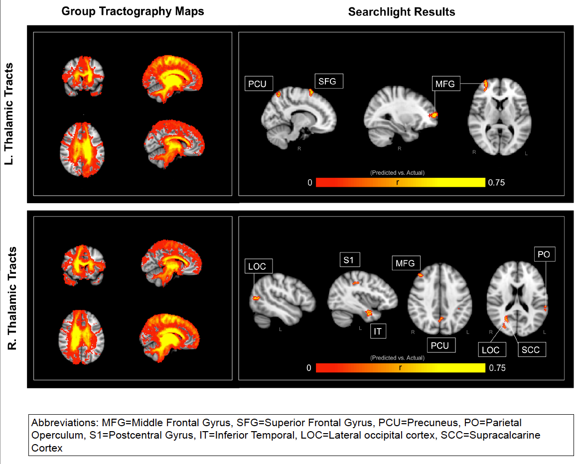
Brief: In this study we used probabilist tractography on diffusion tensor imaging data to look at the whole brain’s anatomical connectivity index with the Left and Right Thalamus for patients with varying disorders of consciousness. Using a searchlight mapping procedure, we trained a Support Vector Regression on all but one patient and attempted to predict a left out subject’s “Coma Recovery Scale” value (leave-one-patient-out cross-validation). That is, we looked at each region in the brain and asked the question “Does this region’s connectivity with the thalamus provide us with significant predictive power in regards to a DOC patient’s level of awareness?”. We were able to capture a significant portion of the variance (upwards of 56% of the variance), especially when drawing features for the SVR from regions such as the Middle Frontal Gyrus, Superior Frontal Gyrus, Precuneus, Parietal Operculum, Postcentral Gyrus, Inferior Temporal, Lateral Ocipital cortex, and Supracalcarine Cortex. Of particular intrigue is the contra-lateral importance of these connections. Thus, the ability for the Thalamus to anatomically connect with these regions significantly predicts levels of consciousness.
Highlighted Figure:
SfN 2014 Abstract:
A reliable neural biomarker would serve as a valuable prognostic indicator for the assessment of awareness in patients with disorders of consciousness (DOC). Previous research has suggested that DOC may reflect disconnections in the thalamocortical networks. In this current study, we used probabilistic tractography to investigate the structural connectivity between the thalamus and the rest of the brain in 23 patients with varying levels of awareness as measured by the coma recovery scale-revised (CRS-R). The CRS-R spans six subscales aimed at assessing overt consciousness: auditory, visual, motor, oromotor, communication, and arousal. We obtained a total CRS-R score for each patient by summing across all six-subscale scores, where a maximum total score would be 23 points. We employed a searchlight mapping approach by centering a 5mm sphere at each voxel in the brain. The thalamic-connectivity-index values of voxels within each sphere were used as predictors in a support-vector-regression. The predictive power of our model was assessed by a leave-one-patient-out cross-validation whereby we iteratively trained a support-vector-regression model on 22 subjects and applied that model to the left-out subject. The resulting vector of predicted CRS-R scores correlated with the actual CRS-R scores most strongly when the searchlight was centered in Middle Frontal Gyrus and Precuneus. More specifically, connections with the left thalamus in these regions accounted for upwards of 44% of the variance in CRS-R scores. Furthermore, lateral occipital cortext and Right Supracalcarine Cortex accounted for up to 56% of the variance in CRS-R scores. These results provide neural bases for the level of conscious awareness displayed by DOC patients. More specifically, this investigation highlights the importance of thalamo-prefrontal and thalamo-temporal circuits in establishing a dependable anatomical metric for calculating patients’ CRS-R scores. Such findings support the “disconnection syndrome” hypothesis by illustrating that decreases in structural connectivity throughout the brain correlates with degradations in conscious awareness.
References
1. Schiff, N.D., 2010. Recovery of consciousness after brain inury: a mesocircuit hypothesis.
Trends Neurosci. 33, 1-9.
2. Van der Werf, Y.D., Witter, M.P., Groenewegen, H.J., 2002. The intralaminar and midline
nuclei of the thalamus. Anatomical and functional evidence for participation in processes of
arousal and awareness. Brain Res Brain Res Rev. 39, 107-40.
3. Kim, S.P. Hwang, E., Kang, J.H., Kim, S. & Choi, J.H. Changes in the thalamocortical
connectivity connectivity during anesthesia-induced transitions in consciousness.
Neuroreport 23, 294-8 (2012).
4. Laureys S. et al. Restoration of thalamocortical connectivity after recovery from persistent
vegetative state. Lancet 355, 1790-1 (2000).
5. Lutkenhoff, E.S., McArthur, D.L., Hua, X., Thompson, P.M., Vespa, P.M., & Monti, M.M.
Thalamic atrophy in antero-medial and dorsal nuclei correlates with six-month outcome
after severe brain injury. NeuroImage: Clinical, 3, 396-404. (2013)
